
English 


Views: 0 Author: Site Editor Publish Time: 2026-05-20 Origin: Site


Dealing with chronic canine ear infections feels like an endless, exhausting battle for many dog owners. You clean their ears repeatedly, yet the frantic scratching, head shaking, and foul odors always seem to return. Relying on "blind" cleaning at home severely limits your ability to actually see the root cause. Without proper visibility, you risk pushing waxy debris further down the canal. You might even miss deep-seated inflammation entirely. This blind approach simply masks symptoms rather than solving them.
A specialized Pet Otoscope serves as the crucial diagnostic tool bridging this frustrating gap between guessing and effectively treating. In this guide, we will explain exactly what clinical tools veterinarians use today. We will detail how modern video otoscopy safely navigates complex anatomy. Finally, you will learn how to confidently evaluate consumer-grade devices for safe at-home monitoring.
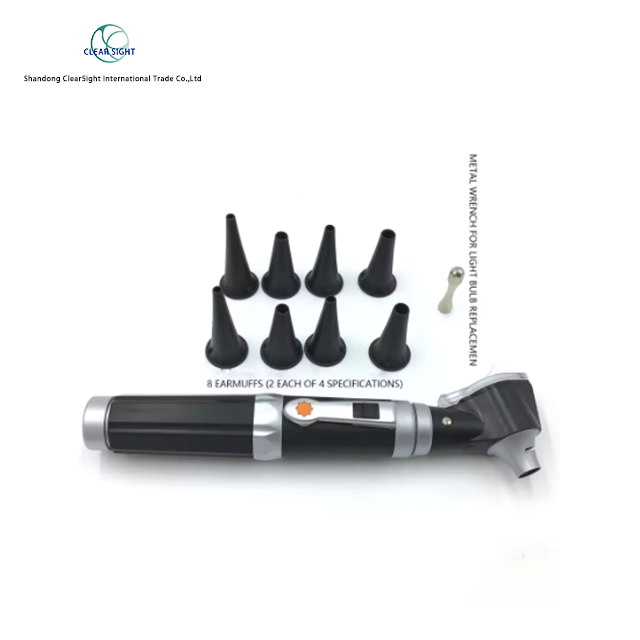
Vets use specialized pet otoscopes designed specifically to navigate a dog’s unique L-shaped ear canal without causing trauma.
Professional clinical diagnostics rely on video otoscopy, which features built-in working channels for deep flushing, biopsy, and exact targeted treatment.
Consumer-grade pet otoscopes offer a high ROI for early detection and remote vet consultations, but cannot replace clinical tools for deep biological removal (e.g., biofilms).
Never use hydrogen peroxide for cleaning; standard monitoring requires specialized tools, and serious interventions often require sedation to ensure patient safety and procedure accuracy.
Veterinary diagnostics require highly specialized tools. Human instruments simply fail when applied to canine anatomy. Understanding this physical barrier helps explain why specialized equipment remains absolutely necessary.
Dogs possess a unique "L-shaped" ear canal. The pathway drops vertically from the outer ear opening. It then makes a sharp, nearly 90-degree turn horizontally toward the eardrum. Standard human otoscopes feature short, wide tips designed for straight canals. Pushing a human tool into a dog's ear causes immediate pain. It also fails to "turn the corner" to reveal the deeper horizontal section.
To safely navigate this bend, veterinarians use a dedicated Pet Otoscope. These devices feature longer, specifically angled attachments designed to bypass the vertical drop safely.
A clinical-grade diagnostic device relies on two primary physical components:
Speculum: This cone-like attachment safely parts sensitive ear tissue. It comes in varying lengths and diameters to accommodate different dog breeds.
Light Source & Optics: High-intensity LED lighting pairs with a powerful magnifying lens. This combination illuminates the dark canal and brings the tympanic membrane (eardrum) into sharp focus.
When professionals look down a dog's ear, they assess several biological markers. They check for erythema, which indicates severe redness and inflammation. They look for active ulcerations along the canal walls. They also monitor for tissue swelling causing a dangerous narrowing of the canal. Finally, they identify specific discharge types. For example, dark, coffee-ground debris strongly indicates the presence of Otodectes cynotis, commonly known as ear mites.
Feature | Human Anatomy & Tools | Canine Anatomy & Tools |
|---|---|---|
Canal Shape | Relatively straight and horizontal. | L-shaped (vertical drop, horizontal turn). |
Speculum Design | Short, wide, and rigid. | Long, narrow, tapered, and sometimes articulating. |
Visual Target | Immediate view of the eardrum. | Requires lifting the pinna to align the dual canals. |
Veterinary dermatology has evolved rapidly over the last two decades. The shift from handheld scopes to digital platforms changed everything. It improved diagnostic accuracy and dramatically increased patient safety.
Older clinical methods relied on basic handheld scopes. This approach forced veterinarians into a dangerous "half-blind" flushing method. Handheld tools keep the light source and magnifying lens too far from the actual tip. This distance makes deep canal assessment incredibly difficult. It also forces the veterinarian into a risky "head-to-head" posture. Placing a doctor's face inches away from an anxious, pained dog creates severe bite risks.
Modern clinics now rely on advanced video otoscopy. These professional-grade systems eliminate blind spots entirely. They utilize high-definition micro-cameras mounted directly at the probe tip. These cameras transmit crisp, magnified images to external wall monitors.
The most critical feature of a professional video system is the built-in working channel. This hollow tube runs alongside the camera lens. It allows veterinarians to pass specialized micro-instruments directly to the disease site.
Key Clinical Outcomes Achieved:
Precise Vacuum-Drying: Vets suction out excess fluids to prevent secondary yeast infections.
Targeted Biofilm Removal: Vets physically scrub away protective bacterial barriers formed by dangerous pathogens like Pseudomonas.
Complex Micro-Surgery: Vets perform myringotomies (deliberate eardrum incisions) to flush and treat middle-ear diseases safely.
Many pet owners feel hesitant when a vet suggests sedation for an ear exam. It often feels like an unnecessary upsell. However, modern veterinary standards require it for very specific medical reasons. We must frame this practice as a baseline for safety and precision.
Explaining the "why" behind sedation builds trust. A thorough deep-ear exam involves rigid instruments, flushing fluids, and micro-tools. You cannot perform these actions safely on an awake, nervous animal.
Inflamed ears become hyper-sensitive to the slightest touch. Dogs will not remain completely still when experiencing this intense pain. They instinctively thrash, shake their heads, or pull away. A moving target makes it physically impossible to safely navigate the L-shaped canal. If a dog jerks unexpectedly, a rigid speculum can easily puncture the delicate eardrum. Sedation eliminates this unpredictable movement entirely.
Effective treatment goes beyond merely looking inside the ear. Deep ear flushing, also known as irrigation, requires high-pressure saline. This process breaks up compacted wax plugs and thick biological debris. Afterward, the vet must safely vacuum-dry the entire canal. Leaving moisture behind fuels explosive bacterial growth. These aggressive, noisy, and uncomfortable procedures are only feasible when the animal is fully sedated and pain-free.
Home monitoring technology has advanced significantly. Pet owners can now purchase consumer-grade devices safely designed for dogs. Evaluating these options requires understanding both the financial benefits and the technical specifications.
Frame the purchase of an at-home Pet Otoscope as a powerful cost-saving measure. Catching minor inflammation early prevents massive medical bills later. Chronic, unchecked infections eventually scar and permanently narrow the ear canal. This irreversible damage often requires Total Ear Canal Ablation (TECA). TECA is a radical surgery removing the entire ear canal, costing thousands of dollars. Early detection stops this painful progression.
When shopping for a device, you must compare specific technological capabilities. Not all tools offer the necessary utility for safe canine use.
Simple vs. Video: Basic lighted magnifiers only work well for superficial, outer-ear checks. Digital models offer Wi-Fi or USB connectivity. This connectivity remains essential for capturing clear images. You can then email these photos directly to your vet for rapid telehealth consultations.
Camera & Lens Specs: Look for specific hardware features. A 140° wide-angle view captures more surrounding tissue. High resolution, such as 1000x1000 pixels or higher, prevents blurry images. Flexible or articulating probes bending up to 275° easily accommodate the L-shape without scraping the walls.
Battery & Portability: A squirmy dog requires rapid, cord-free handling. Seek models featuring rechargeable capacities around 5000mAh for reliable, continuous use.
Device Type | Best Use Case | Data Capture | Cost Range |
|---|---|---|---|
Basic Lighted Speculum | Quick outer-canal checks; checking for large debris. | None (Visual only) | Low |
Digital Wi-Fi/USB Camera | Telehealth consults; deep L-canal monitoring. | HD Photos / Video | Medium - High |
Articulating Endoscope | Navigating complex bends safely in large breeds. | HD Photos / Video | High |
Buying the right equipment solves only half the problem. You must also implement safe handling techniques. User-inflicted trauma occurs frequently when owners rush the process or use improper angles.
Follow strict step-by-step guidance to avoid scraping the sensitive canal lining. Proper preparation ensures a calm dog and a clear visual field.
Begin by cleaning the outer ear (pinna) using a vet-approved flush. Gently wipe away loose dirt and wax using a cotton ball.
Crucial Warning: Never use hydrogen peroxide in a dog's ear. It acts as a harsh oxidizing agent. It damages healthy epithelial tissue and delays healing. Furthermore, it leaves the deep canal dangerously moist. Excess moisture creates the perfect breeding ground for worsened yeast and bacterial infections.
You must align the vertical and horizontal canals manually before inserting any tool.
Position your dog comfortably, ideally sitting or lying down.
Use your non-dominant hand to gently grasp the ear flap (pinna).
Lift the ear flap upward and pull slightly outward away from the head. This action straightens the L-shaped bend.
Take the otoscope in your dominant hand.
Slowly introduce the speculum into the vertical canal, pointing slightly downward and inward.
Always move slowly. You should eventually see a flat, grayish, semi-transparent membrane. This is the eardrum. Stop immediately if you meet any physical resistance. Never force the tool deeper. If the canal appears swollen shut, withdraw the device and call your clinic.
Consumer tools empower pet owners, but they do not replace veterinary medicine. You must establish strict success criteria for home use to avoid delaying necessary medical treatment.
Use your at-home device solely for three specific purposes. First, conduct routine weekly monitoring on healthy ears. Second, perform post-treatment follow-ups to ensure prescribed drops actually work. Third, gather high-quality photo evidence for your veterinarian.
Certain symptoms demand immediate professional intervention. Stop your DIY exam instantly if you observe any of the following red flags:
Pain Responses: Sudden vocalization, yelping, or violent flinching upon touching the outer ear.
Visual Trauma: Clear visual evidence of active bleeding or severe ulceration along the canal walls.
Foreign Bodies: Plant materials, like foxtails or grass seeds, lodged deeply in the canal. Do not attempt to pull these out yourself.
Abnormal Odors: Persistent foul odors emanating from the ear. Sweet smells often indicate heavy Pseudomonas bacterial loads. Vinegar-like smells strongly point to severe yeast overgrowth.
Veterinary dermatologists rely on a simple threshold. If your dog requires ear cleaning more than once a week to stay comfortable, a deeper problem exists. High-frequency cleaning indicates underlying environmental allergies, food sensitivities, or chronic disease. These complex conditions require professional video otoscopy, laboratory cultures, and prescription medications.
Understanding canine ear anatomy fundamentally changes how you manage recurring infections. Veterinarians rely heavily on high-tech video otoscopy systems for micro-surgical treatments, biofilm removal, and deep flushing. However, the basic Pet Otoscope remains an invaluable first-line tool for everyday pet owners.
We highly recommend investing in a digital, consumer-grade device featuring a built-in camera. This technology empowers you to monitor treatments effectively and catch minor inflammation early. Capturing precise photos allows for rapid telehealth intervention. Ultimately, taking a proactive, visually guided approach prevents irreversible ear damage and protects your dog's long-term comfort.
A: Yes. Chronic, untreated bacterial or yeast infections build severe pressure deep inside the ear. This pressure can easily rupture the delicate tympanic membrane. Once ruptured, blind at-home flushing becomes extremely dangerous, as fluids can enter the middle ear and cause neurological damage or deafness.
A: No. You should never use hydrogen peroxide. It damages healthy ear tissue and delays natural healing. It also fails to effectively clear waxy yeast deposits. Worse, it leaves behind excess moisture in the canal, which actively fuels further bacterial and yeast growth.
A: Inflamed ears cause intense pain. Dogs instinctively shake or pull away when touched. Sedation prevents this pain-induced movement. Absolute stillness ensures the vet can safely navigate the complex L-shaped canal with rigid tools without accidentally rupturing the eardrum or scraping the walls.
A: A perfectly healthy canine eardrum appears flat, semi-transparent, and pale gray or pearly white. You should see no outward bulging, no fluid trapped behind the membrane, and no severe redness or vascular swelling along the surrounding canal walls.


