
English 


Views: 0 Author: Site Editor Publish Time: 2026-05-19 Origin: Site


Dealing with chronic canine ear issues is an incredibly frustrating experience for any dog owner. Watching your pet constantly scratch, furiously shake their head, or emit a foul odor creates intense anxiety. You often find yourself wondering if they just have a minor itch or a painful deep infection. The main diagnostic barrier here is your dog's actual ear anatomy. You cannot accurately diagnose medical conditions you simply cannot see from the outside. Guessing the root cause frequently leads to ineffective home treatments or rapidly worsening conditions.
Fortunately, modern diagnostic tools help shift pet care from blind guesswork to clear visual verification. Devices like a high-quality Pet Otoscope allow you to safely examine the ear canal in real-time. They effectively bridge the vital gap between at-home symptom monitoring and professional veterinary intervention, empowering you to make informed healthcare decisions.
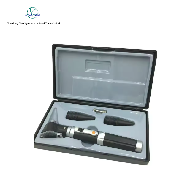
Anatomy dictates visibility: A dog’s “L-shaped” ear canal hides the root causes of infections, making naked-eye inspections ineffective.
Symptoms indicate location: Outer ear issues present as redness and odor, while middle/inner ear problems trigger neurological signs (loss of balance, head tilt).
Tech-enabled monitoring: Utilizing a digital Pet Otoscope allows owners to safely visualize the canal, share images with vets, and verify treatment success.
Safety first: Never use Q-tips, alcohol, or hydrogen peroxide; diagnostic tools must be used solely for observation, not deep intervention.
ROI of prevention: Catching issues early and monitoring medication compliance visually reduces the long-term veterinary costs of chronic, recurring infections.
Dogs possess a unique and complex ear anatomy. Their ear canals form a distinct "L" shape. A steep vertical drop goes down before making a sharp horizontal turn toward the eardrum. This anatomical quirk creates a dark, warm environment. Moisture easily gets trapped inside after a bath or a long swim. Yeast and bacteria thrive rapidly in these damp conditions. Foreign objects frequently become lodged deep inside as well. Grass seeds, for example, are notorious for slipping down the vertical canal and refusing to come out.
Because of this specific shape, naked-eye inspections are essentially useless. Looking at the outer ear flap, known as the pinna, only reveals secondary inflammation. You might notice redness or swelling on the surface. However, the primary trigger usually hides deep within the horizontal canal near the tympanic membrane. You cannot see the eardrum without specialized optical equipment.
Guessing the underlying cause carries significant medical risks. Many owners buy over-the-counter ear drops and blindly squirt them inside. This approach is highly dangerous. If the eardrum is already ruptured, introducing liquids can cause permanent deafness. Severe neurological damage may also occur. Applying the wrong medication often makes the infection much worse. It delays proper healing and prolongs your dog's pain.
Pushing debris further down the horizontal canal worsens the blockage.
Using acidic cleaners on a ruptured eardrum causes excruciating pain.
Failing to identify embedded foreign bodies requires surgical removal later.
Promoting antibiotic resistance happens when owners apply incorrect medicines.
Ear infections do not all look or act the same. Veterinarians categorize these issues based on their specific physical location. Understanding these clinical categories helps you communicate better during a clinic visit.
Outer ear infections are known medically as otitis externa. These issues primarily affect the visible ear flap and the vertical canal. You will easily spot strong visual and olfactory cues here. Dark brown or mucus-like discharge is incredibly common. Sometimes, you might notice crusty debris resembling coffee grounds. This specific texture strongly indicates an active ear mite infestation. Redness, severe swelling, and a pungent, foul odor are also hallmark signs of otitis externa.
Infections can travel deeper, causing otitis media or otitis interna. These conditions affect the delicate middle and inner ear structures. When an infection reaches this depth, it becomes a severe medical emergency. The disease often escalates into vestibular disease. This condition directly affects your dog's sense of balance and coordination.
Red-flag emergency symptoms include:
A pronounced head tilt localized to one side.
Asymmetrical pupil size, where one pupil appears much larger than the other.
Rapid, flickering, involuntary eye movements known as nystagmus.
A sudden loss of balance, stumbling, or continuously walking in circles.
Various everyday factors trigger these painful symptoms. Environmental allergies to pollen frequently disrupt the delicate skin barrier. Moisture retention from frequent swimming encourages aggressive yeast overgrowth. Parasites quickly colonize unprotected ear canals. Underlying systemic issues, like hypothyroidism, can also make certain dogs more susceptible to chronic, lifelong infections.
Condition | Affected Anatomical Area | Common Observable Symptoms | Primary Triggers |
|---|---|---|---|
Otitis Externa | Outer canal & pinna | Redness, foul odor, coffee-ground debris, intense scratching | Allergies, moisture, ear mites, wax buildup |
Otitis Media | Middle ear (behind eardrum) | Pain when opening mouth, lethargy, deep purulent discharge | Untreated outer infections, ruptured tympanic membrane |
Otitis Interna | Inner ear & associated nerves | Head tilt, nystagmus, severe loss of balance | Severe deep infection, internal tumors, polyps |
The traditional approach to canine ear care is inherently reactive. Most pet owners wait until symptoms become severe before taking action. Your dog cries out in pain, so you hastily book an expensive emergency vet visit. The vet examines the ear using a clinical-grade scope. They prescribe a standard round of medication. You apply the drops at home and simply hope the infection clears up. This cycle is stressful, highly expensive, and largely based on blind faith during the recovery period.
A modern approach focuses on proactive, visual monitoring. Today, you can use a consumer-grade Pet Otoscope right in your living room. This device fundamentally changes how we manage canine ear health long-term.
It is vital to understand the proper role of this technology. An at-home camera does not replace a trained, licensed veterinarian. Instead, it serves as an excellent triage and treatment compliance tool. You can capture clear images and video of the ear canal on demand. These visuals help you differentiate between mild, normal wax buildup and active, weeping pus. If you notice concerning discharge, you can email these high-resolution images directly to your clinic. This capability makes telehealth consultations highly effective and ensures you seek professional hands-on help exactly when needed.
Not all digital cameras are suitable for unique canine anatomy. You must carefully evaluate several technical features before purchasing an inspection tool for your dog.
When selecting a Pet Otoscope, prioritize the lens and its focal length. You need a specialized macro lens calibrated specifically for veterinary use. Standard human otoscopes often lack the correct focal distance for an animal's ear. A proper pet lens ensures clear focus in tight, dark spaces. It allows you to see the horizontal canal clearly without pushing the device dangerously deep.
Illumination serves as another critical factor. The canine ear canal is pitch black. Look for a device featuring adjustable LED ring lights. Bright, even lighting penetrates thick, dark discharge effectively. However, the LEDs must remain cool during operation. Hot bulbs can cause severe thermal discomfort or accidentally burn delicate ear tissues.
Form factor and connectivity directly impact everyday usability. You will generally choose between wired and wireless models. Wireless Wi-Fi models are highly recommended for pet owners. Dogs rarely sit perfectly still during an ear exam. They squirm, pant, and aggressively shake their heads. Maneuvering a tethered, wired device around a moving animal is frustrating and totally impractical. A wireless camera paired directly with a smartphone app offers the necessary freedom of movement.
Finally, ensure the device supports high-definition image capture. Your primary goal is veterinary sharing. You need a tool capable of recording crisp photos and HD video. Grainy, low-resolution pictures will not help your vet make an accurate telehealth assessment. High-quality visual evidence streamlines clinic communication immensely.
Owning the right tool is only half the battle. You must execute the physical examination safely to avoid harming your pet. Proper positioning is the most important preparatory step.
To begin, have your dog sit calmly in a quiet, well-lit room. Gently grasp the tip of the ear flap. Pull the flap firmly but gently upward and slightly back toward the dog's tail. This specific action helps straighten out the vertical portion of the L-shaped canal. It creates a much clearer, safer pathway for the camera lens to descend.
Strict risk mitigation rules apply whenever you examine the ear. Below are critical safety guidelines you must follow:
Never force the camera past any physical resistance or inflamed, swollen tissue.
Never use the otoscope tip as a scoop to dig out hardened wax or debris.
Never insert standard cotton Q-tips into the canal. They pack debris deeper and pose a massive eardrum rupture risk.
Never pour harsh household chemicals like hydrogen peroxide or rubbing alcohol into the ear. They destroy healthy tissue and cause excruciating pain.
Sanitization represents another vital safety protocol. Infections easily transfer from an infected ear to a healthy one. You must clean the camera tip thoroughly between uses. Wipe the lens and surrounding casing down using medical-grade alcohol wipes. Allow the device to dry completely before inspecting the second ear. This simple, reliable habit prevents cross-contamination and keeps your equipment completely sterile.
Treating chronic ear infections is undeniably expensive. Consulting fees, cytology swabs, and prescription medications add up very quickly. Understanding the total cost of ownership (TCO) for your pet's healthcare is essential for smart budgeting.
The highest hidden cost comes from treatment non-compliance. Ear infections often look perfectly healed on the outside long before they are actually cured internally. The visible swelling goes down, and the bad odor fades away. Because of this, owners frequently stop applying prescribed drops a few days too early. This mistake leaves microscopic bacteria surviving deep in the horizontal canal. These surviving pathogens breed rapidly. They soon mutate into antibiotic-resistant strains, causing severe, recurring infections that are incredibly difficult to cure.
Veterinarians naturally require follow-up visits to prevent this exact scenario. You pay a secondary exam fee just so the vet can look inside and confirm the infection is gone. These repeated check-ups drain your wallet continuously over the dog's lifespan.
This dynamic highlights the immense return on investment (ROI) of a personal pet inspection camera. Frame the initial upfront purchase price against the long-term savings of preventive care.
You easily avoid paying for unnecessary follow-up visits simply to verify healing.
You prevent the massive veterinary costs associated with treating antibiotic-resistant superbugs.
You catch new flare-ups weeks before they escalate into an expensive emergency room visit.
The device grants you ultimate visual confirmation. You can physically see that the deep canal is truly clear, clean, and pink before you decide to stop the vet-prescribed treatments.
Taking control of your dog's auditory health requires a smart balance of professional medical guidance and diligent home care. While only a licensed veterinarian can accurately prescribe the right antibiotics, antifungals, or anti-parasitic treatments, your daily involvement remains undeniably crucial. Your ability to safely monitor the ear canal directly dictates the long-term success of any medical treatment plan.
Establish a baseline: Inspect your dog’s ears while they are perfectly healthy to understand what normal, pink tissue looks like.
Document everything: Snap photos of any unusual redness or discharge to build an accurate visual history for your clinic.
Upgrade your toolkit: Invest in a specialized smart camera to eliminate guesswork entirely and prevent dangerous home remedies.
Follow through on medication: Visually verify the complete eradication of debris deep inside before you consider stopping any prescribed healing drops.
A: A digital camera can reveal debris visually consistent with specific infections. You might see dark, coffee-ground-like specks indicating mites, or brown, greasy discharge suggesting a yeast overgrowth. However, it cannot provide a definitive diagnosis. Your veterinarian still needs to perform a microscopic swab test, known as cytology, to confirm the exact pathogen before prescribing targeted medication.
A: Yes, it is perfectly safe as long as you follow strict precautions. You must use the device solely for visual inspection within the outer and vertical portions of the canal. Never force the camera tip past any physical resistance or swollen tissue, and never use the lens to aggressively dig out earwax.
A: An ear issue becomes a true medical emergency when it presents with neurological symptoms. If your dog exhibits a sudden head tilt, begins walking in continuous circles, shows unequal pupil sizes, or experiences severe lethargy and extreme pain upon opening their mouth, seek veterinary care immediately.
A: Use a high-quality, vet-approved liquid cleanser designed specifically for canines. Fill the ear canal with the solution, gently massage the base of the ear to break up debris, and let your dog vigorously shake its head. Finally, wipe only the visible outer flap using a soft cotton ball or gauze pad.


